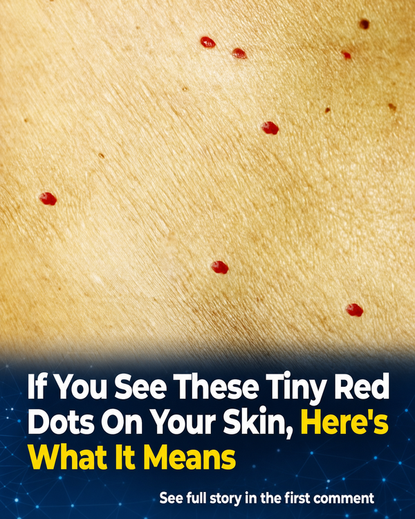
A gradual, stable red speck that hasn’t changed in weeks or months usually points toward something benign. A sudden cluster that spreads over a day or two, particularly on the lower legs, raises different questions and may call for prompt evaluation rather than a wait-and-see approach.
Why Red Dots Appear on the Skin
Dense capillaries sit close to the skin’s surface. When a small group of vessels widens, twists, or grows, it can create a bright pinpoint spot. Blood can also leak outside a vessel entirely, and that mechanism changes the urgency of the situation.
Dermatologists lean on history to sort things out. They ask about recent viral illness, new medications, blood thinners, steroid creams, and skin trauma, since these can thin skin or increase bruising. They also check whether spots itch, sting, or cluster around hair follicles, since folliculitis can mimic a vascular lesion.
Cherry Angiomas: The Common Harmless Culprit
Dr. Haas points to the explanation seen most often in daily practice. He notes that cherry angiomas are what clinicians see most commonly, especially in patients with lighter skin tones. These are benign overgrowths of small blood vessels that often start flat and slowly become small domes, sometimes bleeding if scraped.
They typically appear on the torso, upper arms, and thighs, and tend to multiply with age. Cleveland Clinic estimates that roughly half of adults have cherry angiomas after age 30, and that they become common by age 75. The clinic describes them plainly as harmless red bumps.
Cherry angiomas tend to look uniform, round, and sharply defined. If a spot looks irregular, scaly, tender, or grows rapidly instead, a dermatologist may consider other possibilities, including pyogenic granuloma or an early skin cancer presenting as a pink-red bump.
Removal is possible when a lesion bleeds often or bothers someone cosmetically, but Cleveland Clinic warns that removal can cause scarring and should never be attempted at home, since cutting or tying one off risks infection and uncontrolled bleeding. In-office removal by cautery or laser, with proper wound care, carries far less risk.
Telangiectasia and Sun-Damaged Vessels
Some red dots aren’t dots at all up close, but widened vessels near the surface that form thin lines or a tiny red star shape. This umbrella term, telangiectasia, describes visible dilated capillaries that often show up on the nose, cheeks, and upper chest after years of sun exposure.
Heat, alcohol, topical steroid overuse, and rosacea can all worsen the appearance of these vessels. DermNet NZ describes telangiectasia as visible small linear blood vessels, sometimes called broken capillaries, though in most cases the vessel wall hasn’t actually ruptured; it has simply dilated and become easier to see through thin skin.
Dermatologists often distinguish these vascular marks from bleeding under the skin using a blanching test, or diascopy. As the Merck Manual explains, pressing a slide against a lesion shows whether it lightens under pressure. Vascular marks like telangiectasia typically blanch, while petechiae usually do not.
Treatment is optional. Many people leave telangiectasia alone, but vascular lasers or intense pulsed light can reduce their appearance when desired. Daily broad-spectrum sunscreen and gentle, non-irritating skin care can help limit new visible vessels over time, particularly on sun-exposed areas.
Petechiae, Purpura, and When Red Dots Signal Urgency
Petechiae look like tiny red or purple pinpoints, generally under 2 millimeters according to StatPearls, and they often cluster on the lower legs and ankles where gravity increases pressure. Unlike cherry angiomas, they don’t rise above the skin, and unlike telangiectasia, they don’t blanch with pressure because blood has already leaked into the skin.
MedlinePlus notes that areas of bleeding into the skin stay the same color when pressed. Petechiae can follow a clear, benign trigger, such as forceful coughing, vomiting, heavy lifting, or an intense workout, and in those cases they often fade within days without other symptoms.
But petechiae can also signal a platelet problem. Platelets help blood clot, so a low count can cause this kind of pinpoint bleeding. The NHLBI lists petechiae as a sign of immune thrombocytopenia, describing them as small, flat red spots caused by blood leaking from vessels under the skin.
Cleveland Clinic advises seeking medical attention if petechiae spread quickly or appear alongside other symptoms. The CDC notes that meningococcal disease can present with a petechial or purpuric rash together with sudden fever, an urgent combination that shouldn’t be ignored. Mayo Clinic similarly advises seeing a healthcare provider if petechiae appear all over the body or the cause can’t be identified.
What a Dermatologist Checks—and When to Rule Out Skin Cancer
Dermatologists don’t treat every red dot as cancer, but they do treat uncertainty as a reason to examine closely. They look at whether a spot blanches, whether it has scale or crust, whether it bleeds easily, and how long it has been present.
Because melanoma and other skin cancers can appear as a new pink or red spot rather than a dark mole, new or changing lesions on sun-exposed areas like the face, scalp, ears, chest, and forearms deserve attention, especially with repeated crusting after apparent healing.
Dermoscopy can help distinguish vessel patterns that suggest a benign angioma from those in a suspicious lesion. When uncertainty remains, the American Academy of Dermatology notes that a biopsy is the only way to know for certain whether a spot is skin cancer.
At home, tracking spots with photos over time, checking whether a spot blanches under gentle pressure with a glass, and noting location, itching, bleeding, or symptoms like fever, mouth bleeding, or confusion can help decide whether to simply monitor or seek prompt care. Bringing a medication list, including supplements, to any appointment can also help a clinician sort a cosmetic spot from a warning sign more quickly.